 By Ava Walters
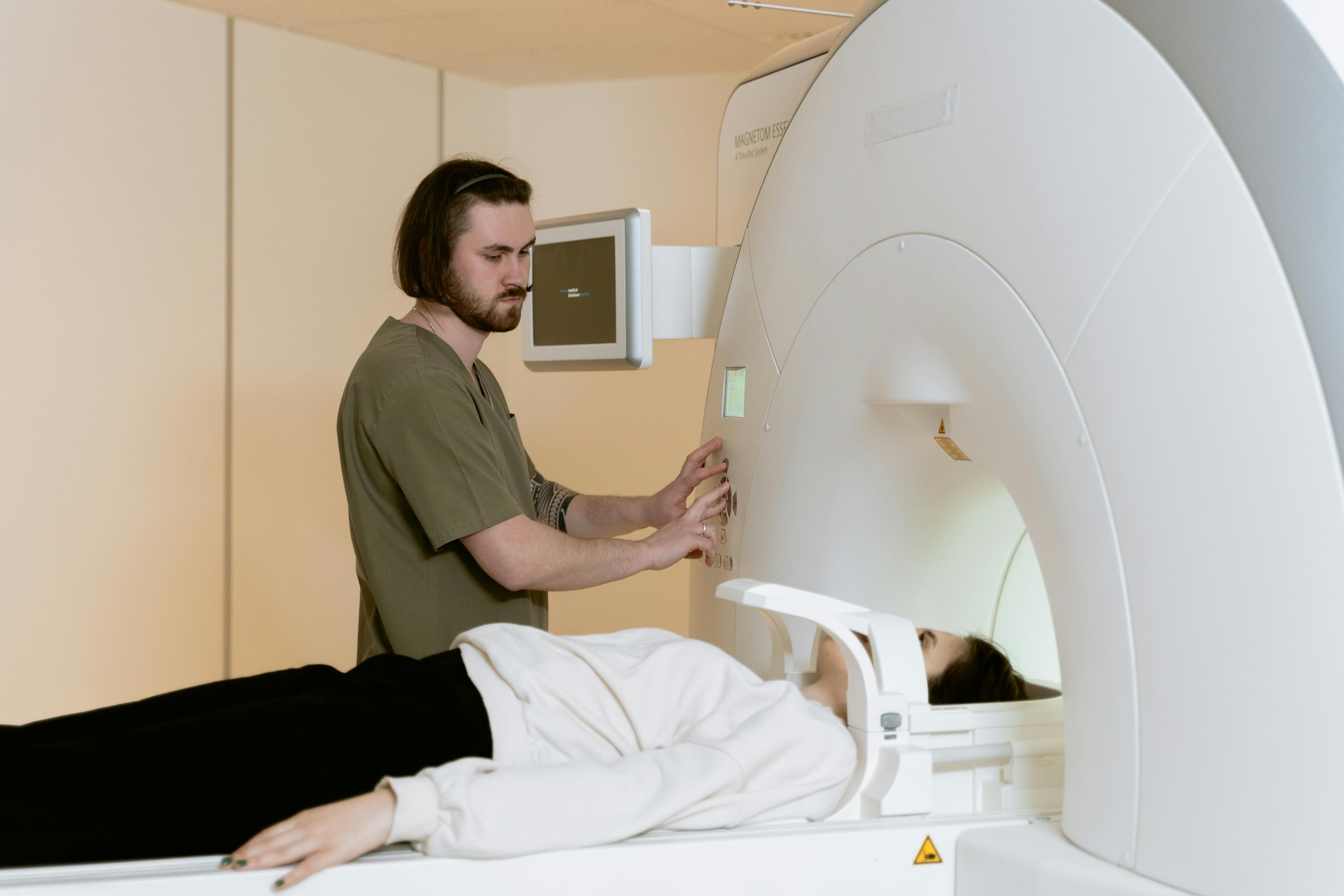
Nuclear medicine scans are a crucial element of modern medical diagnostics, offering detailed insights into the physiological processes occurring within the body. Unlike traditional imaging techniques that primarily focus on structural aspects, nuclear medicine provides functional information by using small amounts of radioactive materials called radiotracers. These scans are pivotal in diagnosing and managing a variety of conditions, from cancer and heart disease to neurological disorders. Understanding the nuances of nuclear medicine scans can help patients and healthcare providers make informed decisions regarding diagnostic and treatment strategies.
What are Nuclear Medicine Scans?
Nuclear medicine scans involve the use of radiotracers, which emit gamma rays detectable by special cameras. These scans are distinct from other medical imaging services because they provide information about how tissues and organs function, rather than just their structure.
- Radiotracers: Administered intravenously, orally, or by inhalation, depending on the type of scan.
- Gamma Cameras: Capture the emitted energy to create images of the body”?s interior.
- High Sensitivity: Allows for early detection of diseases by identifying changes at the cellular level.
Types of Nuclear Medicine Scans
Various types of scans are utilized depending on the clinical indications. Each type of scan is designed to target specific organs or conditions.
Positron Emission Tomography (PET) Scans
PET scans are widely used in advanced diagnostic imaging to detect cancer, assess heart function, and evaluate brain abnormalities. They are highly effective in:
- Detecting cancerous tissues and monitoring treatment progress.
- Evaluating blood flow to the heart and identifying coronary artery disease.
- Assessing brain disorders like Alzheimer’s disease and epilepsy.
Single Photon Emission Computed Tomography (SPECT) Scans
SPECT scans, a common form of nuclear medicine imaging, are frequently used to diagnose and manage various conditions, including:
- Bone disorders, such as fractures, infections, or tumors.
- Thyroid abnormalities, including hyperthyroidism or thyroid cancer.
- Heart diseases, by evaluating blood flow to heart muscles.
Benefits of Nuclear Medicine Scans
Employing nuclear medicine scans provides several advantages over traditional imaging methods:
- Early Detection: Identifies diseases at an earlier stage, leading to more effective treatment options.
- Functional Imaging: Offers insights into organ function, aiding in accurate diagnosis.
- Non-invasive: Minimizes discomfort as the procedures are generally painless.
Considerations and Safety
While nuclear medicine scans offer numerous benefits, certain considerations must be kept in mind. The use of radioactive materials, although minimal, requires careful handling and expertise. It is recommended to consult with a nuclear medicine clinic for personalized advice and safety guidelines.
- Radiation Exposure: The amount is low and similar to that of traditional X-rays, but not suitable for pregnant women unless absolutely necessary.
- Allergies: Rare, but possible allergic reactions to radiotracers should be discussed with healthcare providers.
- Preparation: Specific instructions may be given, such as fasting or avoiding certain medications.
Conclusion
With the integration of cutting-edge technology, nuclear medicine scans have become an indispensable tool in the realm of radiology imaging services. Their ability to provide functional information about organs and tissues enables healthcare professionals to diagnose and treat a wide range of conditions effectively. As radiological imaging services continue to evolve, the precision and efficacy of nuclear medicine will undoubtedly enhance patient care and outcomes.